Cognitive Control
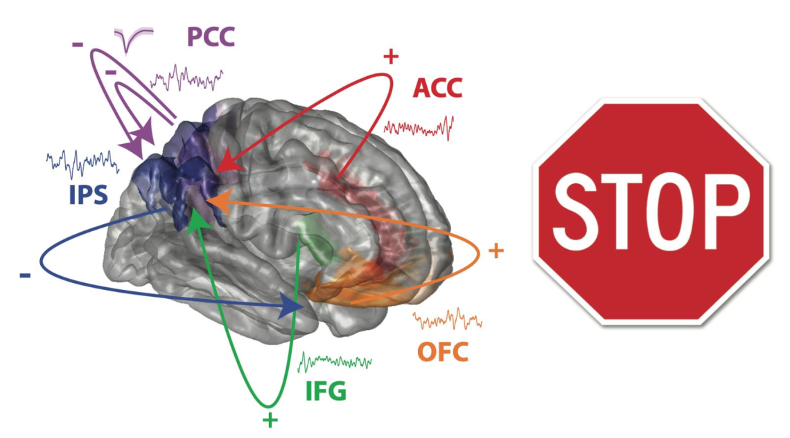
One focus area of the lab is understanding the brain dynamics subtending the ability to adjust and control ongoing behavior to best interact with the environment. Cognitive control mechanisms override habitual responses and allow us to display flexible behaviors. We employ inhibition control paradigms (go/no-go, stop-signal task) while recording local field potentials (LFP) and single-unit activity (SUA) across several cortical areas in human participants. We are particularly interested in understanding the factors that modulate control abilities, such as the state of ongoing neural dynamics and network-level dysfunctions.

Neuromodulation
The lab is dedicated to developing tools to evaluate connectivity across brain regions employing brain stimulation and understanding how to tune brain stimulation protocols to engage specific brain regions and functional networks. In collaboration with Rice University, we developed a biophysical model to estimate brain connectivity based on single-pulse electrical stimulation (SPES) and pulse-evoked potentials (PEP). We combine cognitive experiments with stimulation protocols to alter functional networks and quantify the effect of neuromodulation on cognitive abilities and behavior.

Visual information encoded in brain oscillations
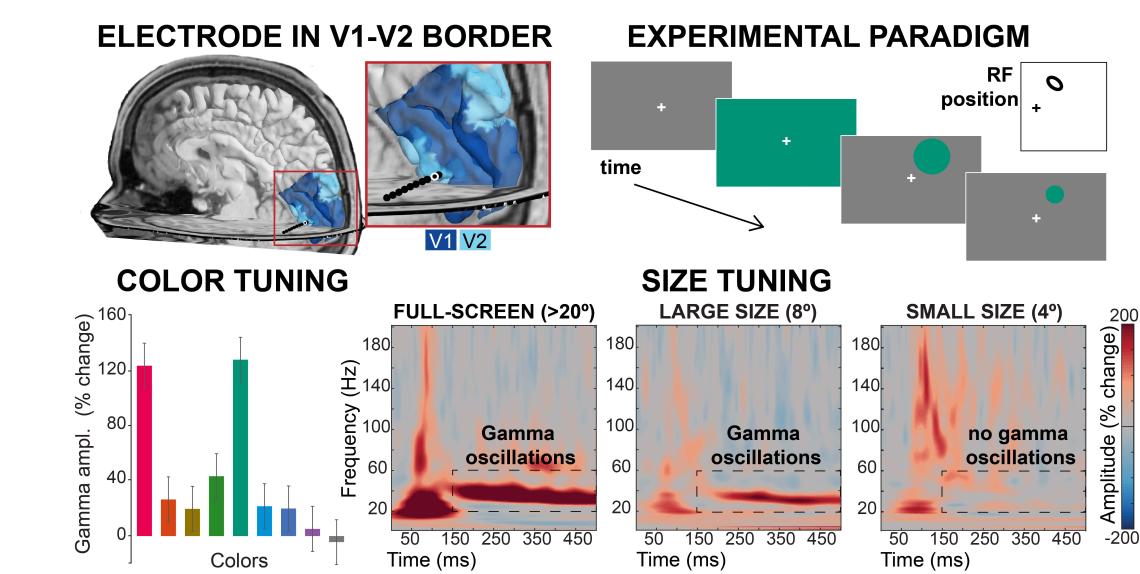
Visual gamma oscillations are high-frequency oscillations occurring in occipital cortex in response to some types of visual stimuli. We are interested in determining the visual features that modulate the presence of these oscillatory patterns, ultimately to decode the information carried by these signals. In collaboration with UPenn, we obtained LFP recordings from occipital and inferior-temporal cortex in human participants during perceptual experiments aimed at isolating different visual features (colors, contrast, visual categories) and we are working on formalizing how gamma oscillations depend on the presence of some visual features (in the visual field) and their position relative to the neural population receptive field.

Brain and cortical parcellation visualization
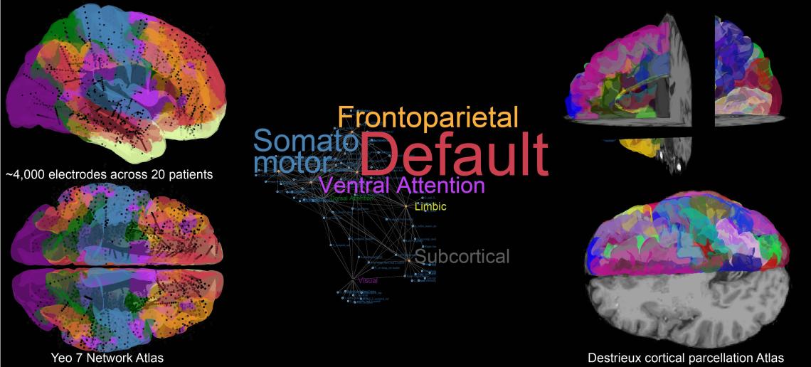
We are always trying to find better ways to visualize and objectively label our recording sites with fine-grained anatomical information combined from multiple sources (i.e., brain atlases, distance from anatomical landmarks, gyral/sulcal geometry, etc.).
Collaborations

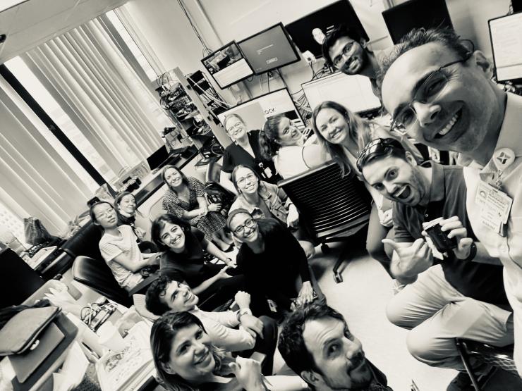
The lab collaborates closely with the whole team performing human intracranial recordings at Baylor College of Medicine: Sameer Sheth and his lab, Kelly Bijanki and her lab, Benjamin Hayden, Andrew Watrous, Nicole Provenza. In addition, we collaborate with Sarah Heilbronner and Jeff Yau. Outside of Baylor College of Medicine, we share projects with Alessandro Alabastri (Rice University), Brett Foster (UPenn), Hernan Rey (Medical College of Wisconsin), Ben Shofty (University of Utah) and Dora Hermes (Mayo Clinic).


