View full size
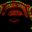
A coronal tissue section cut through the adult cerebellum stained with Phospholipase β4 and Zebrin II to illustrate the complementarity of Purkinje cell stripe patterns.
View full size
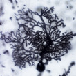
A single Purkinje cell labeled using the classic Golgi-Cox method. The elaborate dendritic tree is revealed with exquisite detail.
View full size
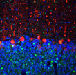
Triple fluorescent labeling of the cerebellum with Calbindin, Cacna2d1 and DAPI.
View full size
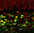
Double immuno fluorescent staining using Calretinin and alpha-Synuclein antibodies to highlight unipolar brush cells and mossy fiber terminals in the granular layer.
View full size
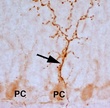
Afferent termination in the rat cerebellum revealed using the expression of cocaine-and amphetamine regulated transcript (CART) peptide.
View full size
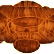
Whole mount Zebrin II peroxidase immunohistochemistry of the mouse cerebellum showing the complex parasagittal pattern of Purkinje cell stripes.


