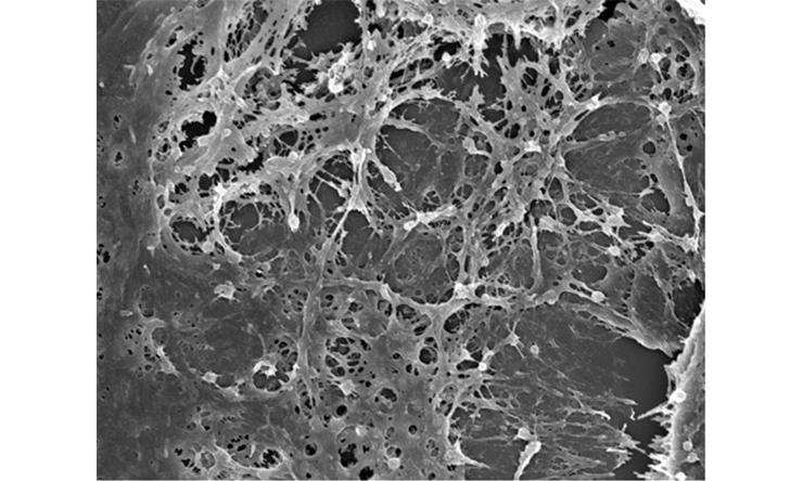
Scanning electron microscopy of extracellular matrix deposited in vitro by mouse cardiac fibroblasts.

Collagen structure in the mouse heart evaluated by the picrosirius red staining under a polarized light.

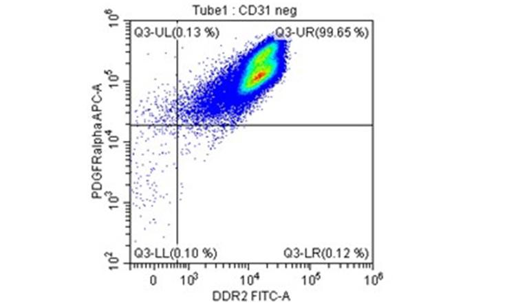
Cardiac fibroblasts isolated from the mouse heart are analyzed by flow cytometry.

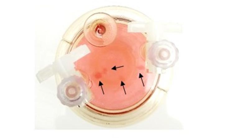
3-D tissue culture with arrows pointing to tissue formation.
About the Lab
Our lab focuses on studying cellular and molecular mechanisms leading to cardiac dysfunction with aging.
Within the last decade, we discovered that the fibroblasts isolated from the old mouse heart have an altered phenotype compared with those from young hearts. In the old heart, fibroblasts become more activated and secrete higher levels of various extracellular matrix components (such as various collagen types and fibronectin containing extra domain A) and inflammatory cytokines such as MCP-1, CX3CL1 and IL-6.
As they produce more matrix components, the heart becomes more fibrotic, which directly leads to diastolic dysfunction. Because of inflammatory chemokines, leukocytes from the blood are chemoattracted into the heart. In the aging heart environment, some of the infiltrating monocytes polarize into pro-fibrotic or pro-inflammatory macrophages.
The lab also examines the possible therapeutic approaches to correct defective pathways and “normalize” pathological phenotypes, and thereby prevent further deterioration of cardiac function.
Lab Projects
Trial J, Heredia CP, Taffet GE, Entman ML, Cieslik KA. Dissecting the role of myeloid and mesenchymal fibroblasts in age-dependent cardiac fibrosis. Basic Research in Cardiology: 2017,112(4):34 PMCID: PMC5591578
Cieslik K.A., Trial J., Entman M.L. Defective myofibroblast formation from mesenchymal stem cells in the aging murine heart. Rescue by activation of the AMPK pathway. American Journal of Pathology 2011, 179, 1792-1806. PMCID: PMC3181380
Lab Location
Physical Lab Location:
One Baylor Plaza
Jones Building
Anderson Hall 540E & 527E
Houston, Texas 77030
Mailing address:
One Baylor Plaza, Mailstation BCM285
Houston, Texas 77030
Administration Contact
Karima Ghazzaly
Manager, Research Administration
Phone: 713-798-4138
Email: hazzaly@bcm.edu
Lab News
Dr. Katarzyna Cieslik was invited to be a member of the National Institutes of Health Cardiac Contractility Heart Failure Study Section for July 2020-June 2024.


