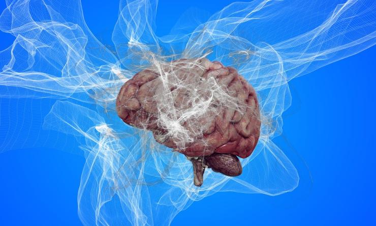
Tau transforms from ‘good guy’ to a ‘bad guy,’ contributes to Alzheimer’s disease
Researchers at Baylor College of Medicine, the Jan and Dan Duncan Neurological Research Institute (Duncan NRI) at Texas Children’s Hospital and collaborating institutions discovered that the enzyme TYK2 transforms the normal protein tau into one that accumulates in the brain and contributes to the development of Alzheimer’s disease in animal models. Published in Nature Neuroscience, the study suggests that partially restraining TYK2 could be a strategy to reduce tau levels and toxicity.
“Many studies have shown that the accumulation of tau in neurons and glial cells in the brain is a main characteristic of Alzheimer’s disease and at least 24 more neurological diseases,” said first author Dr. Ji-Yoen Kim, assistant professor of molecular and human genetics at Baylor in the lab of Dr. Huda Zoghbi. Zoghbi, the corresponding author of the work, is a Distinguished Service Professor at Baylor, director of the Duncan NRI and a Howard Hughes Medical Institute (HHMI) investigator.
Previous studies showed that tau is chemically modified in disease, predominantly by the addition of extra phosphate to the Tyrosine groups in the protein, and that these changes play a crucial role in regulating tau accumulation.
The Zoghbi lab had earlier identified TYK2 – an enzyme that adds phosphate to Tyrosine groups – as a potential regulator of tau levels and that knocking down the TYK2 gene reduced tau levels in human cells. In the current study, the team dug deeper into how TYK2 transforms tau into a protein that aggregates and propagates to neighboring cells and accumulates in tangles inside cells, influencing the development of tau-driven neurodegeneration.
Working with human cells and animal models of tau-driven dementia, the researchers are the first to show that TYK2’s modifications to tau contribute to tau-mediated disease. “We found that TYK2 adds phosphate groups to tau at a particular location on the protein identified as Tyrosine 29,” Kim said. “This modification stabilizes tau levels in human cells and mouse neurons by making it resistant to autophagy, a cellular process important for clearing proteins,” Kim said. “Impervious to clearance, modified tau accumulates in the brain.”
The finding that TYK2 enhances the aggregation of tau suggested that manipulating TYK2 might help regulate tau aggregation and its consequences. The team tested the effect of partially reducing TYK2 in two mouse models and found that this was sufficient to reduce tau levels and mitigate its accumulation. “Although much work is needed, our findings suggest that partial inhibition of TYK2 could thus be a strategy to reduce tau accumulation and toxicity,” Kim said.
“To this end, we are encouraged by the fact that others have developed TYK2 inhibitors that have been tested in humans for other indications,” said Zoghbi. “Studies are needed to see if these inhibitors indeed get into the brain and lower tau levels to explore their potential effects in Alzheimer’s disease and tau-induced dementias.”
Bakhos Tadros, Yan Hong Liang, Youngdoo Kim, Cristian Lasagna-Reeves, Jun Young Sonn, Dah-eun Chloe Chung, Bradley Hyman and David M. Holtzman also contributed to this work. The authors are affiliated with one or more of the following institutions: Baylor College of Medicine, Jan and Dan Duncan Neurological Research Institute at Texas Children’s Hospital, Indiana University School of Medicine, Harvard Medical School and Massachusetts General Hospital, Washington University in St. Louis and Howard Hughes Medical Institute.
This work was funded by JPB Foundation, HHMI, Eunice Kennedy Shriver National Institute of Child Health and Human Development NIH grant P50HD103555 and NIH/NINDS grant R01NS119280 and the Ting Tsung and Wei Fong Chao Foundation.


