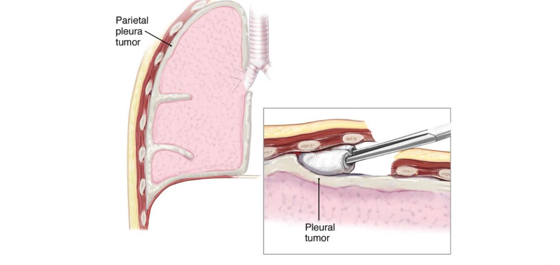
Pleurectomy and decortication is a surgical procedure that may be recommended for patients with mesothelioma or other tumors or diseases affecting the inner chest. This involves removing diseased tissue to alleviate symptoms and improve lung function.
During the procedure, the patient is positioned on their side, and an S-shaped thoracotomy incision is made. The sixth rib is removed to provide access, and the space between the lung and chest cavity is carefully dissected—a process of separating tissues to expose and address affected areas. The tumor is then separated from the chest wall. Once a sufficient area of the chest wall has been cleared, a chest retractor is inserted to facilitate further surgical steps.
The pleura now can be moved from the mediastinum. Once the upper portion of the lung is completely detached from the chest wall, the vessels entering the lung are exposed. On the left side, the esophagus and aorta are identified and dissected. On the right side, the superior vena cava is dissected away from the tissue and tumor being removed. The dissection then continues behind the pericardium.
In some patients, involvement of the diaphragm necessitates a full-thickness resection of the diaphragm muscle.
Once the dissection is completed to expose the vessels connecting the heart and lungs, the space between the lung and chest wall is opened decortication of the visceral pleura (membranous outer layer of the lung) is performed. During decortication, the lung is deflated to reduce blood loss. Most patients require blood transfusion during the procedure. Additionally, lymph node dissection, a process of removing lymph nodes to assess or manage disease, is also performed.
Once the tumor is removed, reconstruction of the pericardium (sac around the heart) and diaphragm, if required, is performed. If the diaphragm is largely intact, it can be pulled downward and to prevent upward movement and improve ventilation.
On the right side, reconstruction of the diaphragm is sometimes performed with a double layer of mesh. On the left, Gore-Tex is used because thicker, non-absorbable material can be required. The prosthesis is secured laterally by placing sutures around the ribs.
An electrocoagulator may be used to help control bleeding from the chest wall. Three chest tubes are placed at the top, and a right-angled tube is placed along the diaphragm to control air leaks that are anticipated and should permit full refilling of the lung.
References
Sugarbaker DJ, Bueno R, Krasna MJ, Mentzer SJ, Zellos L (eds). Adult Chest Surgery. New York: The McGraw-Hill Company, 2009.
Sugarbaker DJ, Norberto JJ, Swanson SJ. Surgical staging and work-up of patients with diffuse malignant pleural mesothelioma. Semin Thorac Cardiovasc Surg. 9:356-60, 1997.
McCormack PM, Nagasaki F, Hilaris BS, Martini N. Surgical treatment of pleural mesothelioma. J Thorac Cardiovasc Surg. 84:834-42, 1982.
Rusch VW. Pleurectomy/decortication in the setting of multimodality treatment for diffuse malignant pleural mesothelioma. Semin Thorac Cardiovasc Surg. 9:367-72, 1997.


 Credit
