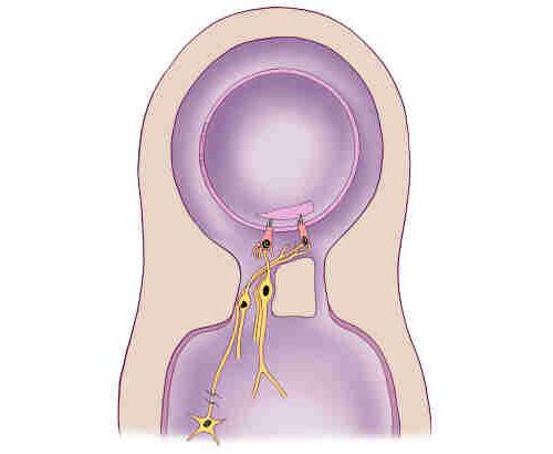
The inner ear is divided into two fluid filled chambers - one inside the other. Figure 3 illustrates the basic organization of both the organs of hearing and balance. The fluid in the two chambers differs on the basis of the kind of salt that each contains. The fluid in the outer or bony chamber is filled with a sodium salt solution (called perilymph) that resembles the salt composition in the blood or the fluids found in the brain. The inner or membranous chamber is filled with a potassium salt solution (endolymph) that resembles the fluid that is normally found inside the cells of the body. The difference in concentration between the two chambers is maintained by specialized cells that line parts of the membranous chamber and "pump" potassium into the membranous chamber. The difference in the chemical composition of the these two fluids provides chemical energy (like a battery) that powers the activities of the sensory cells.
This division of labor is unique to the inner ear because the function of the principal cells relies on chemical energy provided by other cells. In virtually all other systems, whether it is heart muscles, the brain, or the retina of the eye, the principal cells must combine nutrients and oxygen to produce the energy they use to perform their functions. In the inner ear, metabolic processes are performed by an organ called the stria vascularis located a half a millimeter from the hearing organ. The stria vascularis is essentially a battery whose electrical current powers hearing. It is powerful enough that if its power could be harnessed it could used instead of batteries for hearing aids.
Figure 3 illustrates a simplified diagram showing the organization of inner ear organs of hearing and balance. The inner ear contains two fluid chambers, a membranous and a bony chamber. The membranous chamber is filled with endolymph while the bony chamber is filled with perilymph. The wall of the membranous chamber is made up of many cells that are so tightly joined together that they prevent the two fluids from mixing. The sensory epithelium makes up only a small portion of the wall of the membranous chamber and contains sensory receptor cells and surrounding supporting cells (supporting cells are not shown in the drawing). The sensory cells are called hair cells because of their appearance under the microscope. Each hair cell has a tuft of projections at one end that looks like a bad haircut. These tiny "hairs" are called stereocilia and are arranged in rows that increase in length towards one side of the cell. The hair cells are connected to neurons at the other end of the cell away from the stereocilia. Neurons have long projections that conduct electrical signals to the brain. When a hair cell is mechanically stimulated it releases a chemical that modulates the electrical activity of these neurons, thereby signaling the brain. In addition to sending information to the brain, some hair cells receive input from neurons that originate deep in the brain and send one of their long projections to the hair cells. These neurons appear to regulate the sensitivity of the hair cells, in a manner similar to changing the volume control on a radio.
Next chapter: Small Size Means Greater Mechanical Sensitivity
Return to the table of contents of How the Ear Works


