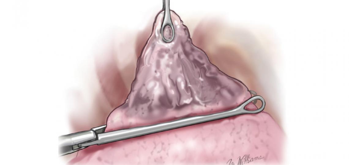
credit: Marcia Williams
A pulmonary bleb is a small collection of air between the lung and the outer surface of the lung (visceral pleura) usually found in the upper lobe of the lung. When a bleb ruptures the air escapes into the chest cavity causing a pneumothorax (air between the lung and chest cavity) which can result in a collapsed lung. If blebs become larger or come together to form a larger cyst, they are called bulla. Unless a pneumothorax occurs, or the bulla becomes very large, there are usually no symptoms. Patients with blebs will typically have emphysema.
Before the surgery, patients are intubated. Patients with emphysema typically have a bronchoscopy procedure before the endotracheal tube is placed. Patients who have underlying emphysema have an epidural catheter placed before surgery and sometimes during the procedure.
Resection
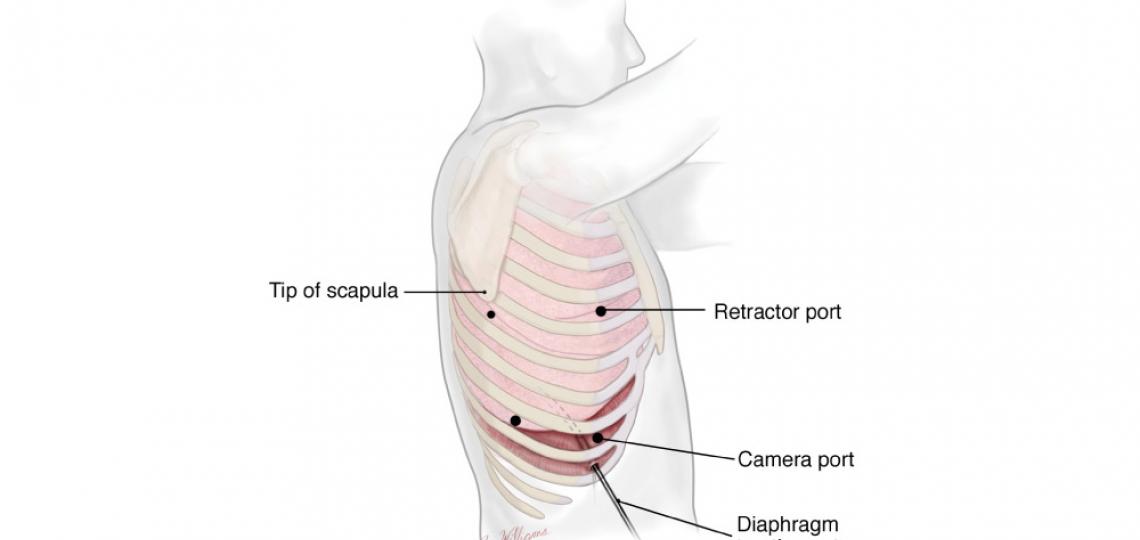
credit: Marcia Williams
The operation for bleb resection can be done via mini-thoracotomy or thoracoscopy. The procedure is performed with general anesthesia using a special endotracheal tube that allows intentional collapse of the lung which is operated on.
The procedure is performed through a series of small incisions. The camera incision is created first, and the thoracoscope (camera) is inserted for inspection of the chest. The bleb is dissected, stapled and the lung tissue is removed from the chest in a specimen bag, allowing more room for the remaining healthy lung to expand.
After Surgery
For the first day or so, a chest tube is left in to help adhere the once deflated lung to the chest wall (pleural symphysis). The tube will be removed if no leak is apparent, and the patient will be sent home. Patients will undergo physical therapy to promote recovery.


 Credit

