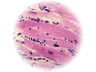
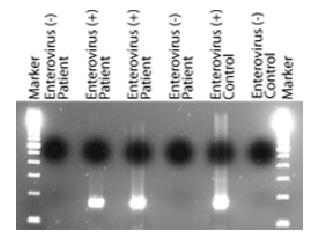
Myocarditis is an inflammatory disorder of the myocardium with necrosis of the myocytes and associated inflammatory infiltrate. It is usually caused by infection with a virus, particularly adenovirus or enterovirus (e.g., Coxsackievirus). These viruses typically cause other less serious diseases, such as gastrointestinal problems and the common cold. Although the utility of myocardial biopsy is debated, suspected myocarditis can be classified based on pathologic findings as defined in the Dallas or Marburg Criteria.
Investigations of the role of viruses in the development of myocarditis, DCM and ARVD/C are ongoing in the Phoebe Willingham Muzzy Pediatric Molecular Cardiology Laboratory, while viral diagnosis is provided by the John Welsh Cardiovascular Diagnostic Laboratory.
Dallas Classification (1987)
Initial Biopsy
- Myocarditis: Myocardial necrosis, degeneration, or both, in the absence of significant coronary artery disease with adjacent inflammatory infiltrate with or without fibrosis
- Borderline myocarditis: Inflammatory infiltrate too sparse or myocyte damage not apparent
- No myocarditis
Subsequent Biopsies
- Ongoing (persistent) myocarditis with or without fibrosis
- Resolving (healing) myocarditis with or without fibrosis
- Resolved (healed) myocarditis with or without fibrosis
WHO Marburg Criteria (1996)
First Biopsy
Acute (active) myocarditis: A clear-cut infiltrate (diffuse, focal or confluent) of >14 leukocytes/mm² (preferably activated T-cells). The amount of the infiltrate should be quantitated by immunohistochemistry. Necrosis or degeneration are compulsory, fibrosis may be absent or present and should be graded.
Chronic myocarditis: An infiltrate of >14 leukocytes/mm² (diffuse, focal or confluent, preferably activated T-cells). Quantification should be made by immunohistochemistry. Necrosis or degeneration are usually not evident, fibrosis may be absent or present and should be graded.
No myocarditis: No infiltrating cells or <14 leukocytes/mm².
Subsequent Biopsies
- Ongoing (persistent) myocarditis: Criteria as in 1 or 2 (features of an acute or chronic myocarditis).
- Resolving (healing) myocarditis: Criteria as in 1 or 2 but the immunological process is sparser than in the first biopsy.
- Resolved (healed) myocarditis: Corresponds to the Dallas classification.
Frequency of Myocarditis

Myocarditis is a rare disease. The World Health Organization reports that incidence of cardiovascular involvement after enteroviral infection is 1-4 percent, depending on the causative organism. Incidence varies greatly among countries and is related to hygiene and socioeconomic conditions. Availability of medical services and immunizations also affect incidence. Occasional epidemics of viral infections have been reported with an associated higher incidence of myocarditis. Studies give a wide spectrum of mortality and morbidity statistics. With suspected Coxsackievirus B, the mortality rate is higher in newborns (75 percent) than in older infants and children (10-25 percent). Complete recovery of ventricular function has been reported in as many as 50 percent of patients. Some patients develop chronic myocarditis (ongoing or resolving) and/or dilated cardiomyopathy and may eventually require cardiac transplantation.
Causes of Myocarditis
Most cases of myocarditis are believed to be caused by virus infection, either directly, due to virus replication, or indirectly, due to autoimmune disease. Occasionally, myocarditis may be due to drug hypersensitivity or toxicity.
Viral Infection

A number of viruses have been identified in the hearts of patients with myocarditis. In 1986 a virus called Coxsackievirus B was identified in the hearts of patients with either myocarditis or DCM. Subsequent work performed by Dr. Jeffrey Towbin and the Muzzy lab has shown that another virus, called adenovirus, is also commonly a cause of these conditions. It has been suggested that when the virus infects the heart it causes the body to react against the virus, much as the body's own defense system (immune system) normally attacks a virus infection, and this causes inflammation in the heart (myocarditis). Usually the virus will be eliminated, however in some patients the immune system may not totally eliminate the virus and in these patients the heart muscle continues to be damaged, leading to DCM. In some people, the body's immune system may attack and damage the heart even though the virus has been eliminated: this is termed auto immune disease.
Auto-Immune Disease
The body's own immune system is responsible for defending it against all foreign invaders for example viruses and bacteria. However, sometimes, this system malfunctions, and starts to attack the body's own tissues - this results in a so-called auto-immune disease. It is thought that this process occurs in some cases of myocarditis.
Other Agents
Myocarditis also can be caused by alcohol, radiation, chemicals (hydrocarbons and arsenic), and drugs.


