Program Overview
Reproductive disorders, contraception, and family planning have major impacts on the health and well-being of individuals world-wide. The goal of this NIH-sponsored training program is to prepare young scientists to address these important problems through basic and translational research and in research-related careers that require expertise in reproductive biology. Baylor College of Medicine is a long-standing leader in reproductive biology research. The goal of our program is to provide the necessary skills and experience to prepare the next generation of scientists for successful and diverse careers in reproductive biology.
Program Design
This program supports two predoctoral and four trainees. The following is a summary of the training program:
- Mentored state-of-the-art biomedical research training in some of the world’s premier reproductive biology laboratories
- Structured committees for both pre- and postdoctoral fellows guided by an Individual Development Plans (IDP) tailored to each trainee’s needs
- Wide-ranging career development activities though the Career Development Center
- Activities to enhance skills in teaching, clinical/translational work, and entrepreneurship within the world's largest medical center
- A mentored journal club that coincides with the Reproductive Biology Lecture series · An annual mini-symposium
- Mentor-mentee training from facilitators trained from the NIH funded National Research Mentor Network
- A novel “Ethics in Reproductive Health Learning Unit”
View a listing of program faculty and program research themes.
Program Research Images

Structure of the Estrogen Receptor (ER)-SRC3-p300 Complex on DNA
The structural model suggests the complex is assembled by each of the two ligand-bound ERα monomers that independently recruits one SRC-3 protein via the transactivation domain of ERα; the two SRC-3s then bind to different regions of one p300 protein through multiple contacts. This was published in Molecular Cell (2015) 57:1047-1058 by Dr. Bert W. O’Malley’s laboratory.

Mouse Sperm Head by Scanning Electron Microscopy
Left, normal sperm head from a control mouse; Right, rounded headed sperm (globozoosperia) from a Ssmem1 knockout mouse. Figure by D. Delvin and K. Nozawa from the Matzuk Lab.

Images from Larina Laboratory
Figure 1: Individual sperm tracking (colored line) toward ovulated eggs within in the mouse oviduct captured in vivo with optical coherence tomography. Image from Shang Wang, PhD, a postdoctoral fellow in the Larina Laboratory.
Figure 2: The mouse female reproduction system imaged with microcomputed tomography (micro-CT) Image by Zheng-Chen Yao in the Larina Lab

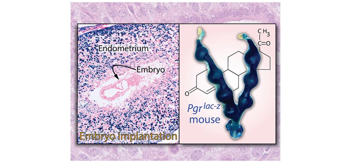
Progesterone Receptor Expression
Visualization of progesterone receptor expression during implantation of a mouse embryo. Left, lacZ stain showing PR expression (blue) and right, whole mount. Images from postdoctoral fellows, Maria M. Szwarc and Lan Hai in the Lydon Lab.

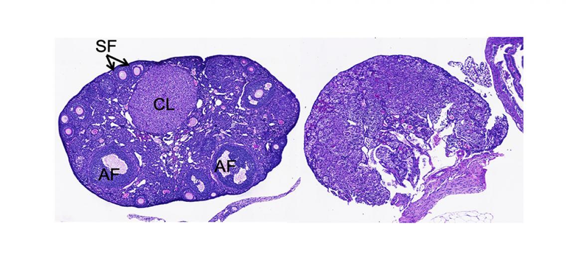
Image from Pangas Laboratory
Ovarian histology from adult wild type (left) and a mouse with oocyte-specific deletion of the SUMOylation enzyme, Ubc9 (right), which are sterile. Photo courtesy of Amanda Rodriguez, a graduate of the Pangas Lab.


