About the Lab
The Wensel Lab research includes the following areas:
- Visual transduction
- G protein pathways and proteins that regulate them in the retina and the brain
- RGS proteins: GAPs for heterotrimeric G proteins
- Biomembranes
- Ocular Proteomics
- Cryo-electron microscopy and tomography
- Gene repair in neurons
- Phosphoinositide signaling
- Super resolution fluorescence microscopy
- Gene engineering in mice and frogs
- TRP Channels
- Primary cilia and retinal ciliopathies
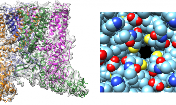
Structure of cation channel TRPV2 in open state

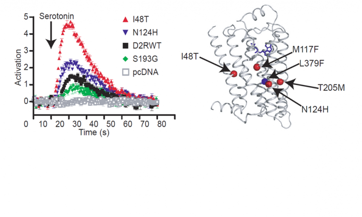
Enhanced Serotonin Responses in Dopamine Receptor Mutants
Serotonin responses of dopamine receptors engineered by evolution-guided mutagenesis.

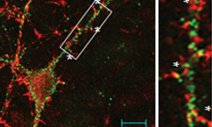
Localization of RGS9-2
Localization of RGS9-2, a regulator of G-protein coupled receptors, in cultured striatal neurons.
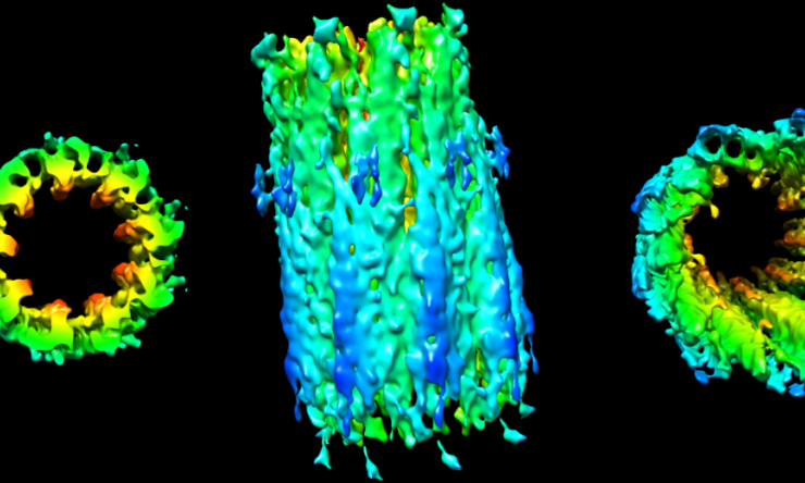
3D structure of daughter centriole by cryo-ET
Cryo-electron tomography and sub-tomogram averaging was used to determine 3D structure of a daughter centriole from a mammalian rod sensory cilium.


 Credit

