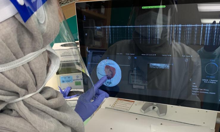
Researchers enrolling patients for study to train AI in assisting breast cancer surgery
The vast majority of breast cancer patients will undergo surgery as part of their treatment, and most of those patients will choose a lumpectomy to remove the tumor and conserve the breast. A surgical oncologist at Baylor College of Medicine is enrolling patients in a study that will use a high-resolution imaging system to collect images of breast tumors in order to develop artificial intelligence. The combination of these technologies currently in development shows promise to help surgeons assess if they have successfully removed an entire tumor.
“One of the big problems in breast cancer surgery is that in about one in four women on whom we do a lumpectomy to remove cancer, we fail to get clear margins. That in turn leads to a need for reoperation to avoid high recurrence rates. Hence the need for a good, effective and user-friendly tool to help us better identify if we have adequately removed the breast cancer from a woman’s breast, to get it right the first time,” said Dr. Alastair Thompson, professor, section chief of breast surgery and Olga Keith Wiess Chair of Surgery at Baylor College of Medicine.
Baylor is partnering with Perimeter Medical Imaging, a medical device company, to use the OTIS™ system, which delivers real-time, ultra-high resolution, sub-surface images of extracted tissues. Perimeter’s AI technology, called ImgAssist, is being developed to overlay OTIS images with areas suspected of containing breast cancer, thus providing another layer of real-time information for surgeons during breast cancer surgery.
“This technique is noninvasive and requires no extra imposition for the patient. OTIS fits into the routine surgical process. The device will examine the tissue sample that is already being extracted,” said Thompson, surgical oncologist at the Dan L Duncan Comprehensive Cancer Center at Baylor St. Luke’s Medical Center and co-director of the Lester and Sue Smith Breast Center at Baylor College of Medicine. “Other imaging technology can be more invasive and may require injections or devices placed in the patient during surgery.”
Perimeter recently received a $7.4 million grant from the Cancer Prevention and Research Institute of Texas (CPRIT) to develop the artificial intelligence algorithm for OTIS using data collected at pathology labs at Baylor College of Medicine, the University of Texas MD Anderson Cancer Center and UT Health San Antonio as part of the ATLAS AI project. Thompson will serve as principal investigator of the study. Enrollment of patients began at Baylor College of Medicine this week. Researchers plan to enroll approximately 400 patients.
Perimeter will continue the ATLAS AI Project with a second randomized, multi-site study in approximately 600 patients to test the OTIS platform with ImgAssist AI against the current standard of care. Researchers will determine whether the platform lowers the re-operation rate for breast conservation surgery.
“This could be a huge improvement for patient care. It could help patients avoid a second surgery and the physical, emotional and financial stress that accompany an additional procedure,” Thompson said.
“Our AI technology has the potential to be a powerful tool for ‘real-time’ margin visualization and assessment that we believe will help physicians improve surgical outcomes for breast cancer patients. The patients who enroll in these clinical studies at Baylor are contributing to new technology that we hope will assist surgeons in the future so that they can reduce the likelihood of their patients needing additional surgeries,” said Andrew Berkeley, co-founder of Perimeter Medical Imaging.
The Baylor study is only open to patients at the Dan L Duncan Comprehensive Cancer Center at Baylor St. Luke’s Medical Center.


