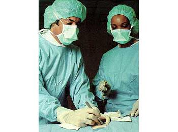
Muscle biopsy is a minor surgical procedure that involves making an incision in the skin to obtain a few small pieces of the underlying muscle for detailed histologic, histochemical, or biochemical examination. The most commonly biopsied muscles are the biceps muscle in the arm and the quadriceps muscle in the leg. Other muscles such as the deltoid and the calf muscles can also be biopsied if indicated.
The procedure is performed under local anesthesia with lidocaine. After the appropriate muscle is selected for biopsy, the area is cleaned with appropriate antiseptic agents and then local anesthetic is applied to the skin and subcutaneous tissue at the site where the incision is to be performed. Usually, the incision is 1.5-2.5 inches long. Once the incision is made, the skin on both sides of the incision is retracted. When the muscle is adequately exposed, sharp scissors are used to remove four to five small pieces of muscle. Local anesthetic is not applied to the muscle, as this would affect the integrity of the tissue and render the histologic examination difficult. Normally, patients report a feeling of mild discomfort or slight and short lasting pain when the muscle is cut. After satisfactory pieces of muscle are obtained, the incision is sutured and a dressing is applied to the area.
The brief discomfort that the patient experiences with the muscle biopsy is related to the injection of the local anesthetic and to the actual cutting of the muscle pieces. When the local anesthetic is injected, the patient feels the needle stick followed by a stinging sensation that lasts a few seconds.
The usefulness of the muscle biopsy depends on issues related to the biopsy itself as well as patient selection. Failure to provide useful diagnostic information all too often reflects improper selection of the biopsy site. When proper standards in muscle biopsy are adhered to, the results are likely to prove valuable. Before a muscle biopsy is obtained, a clinical examination, muscle enzyme determination, and other laboratory tests, including electromyography, are needed. A muscle biopsy is rarely indicated before completing such an evaluation and determining a preliminary diagnosis. The patient must be informed of the reason of the biopsy, choice of biopsy site, technique of biopsy, risks, and possible complications, and the physician's expectation from the study of the biopsy. It is equally important to inform the patient that a muscle biopsy, like all other biopsies, is subject to sampling error and a negative biopsy does not exclude the presence of a suspected disease.
We recommend that muscle biopsies be done by a neuromuscular disease specialist or by a surgeon with experience in obtaining adequate specimens and proper handling of the tissue. If the biopsy has to be done by a surgeon unfamiliar with such matters, it is very important that the surgeon communicates with the neuromuscular specialist or the treating neurologist before the biopsy. Such communication will allow for better biopsy site selection and for proper handling of the tissue after it is obtained.
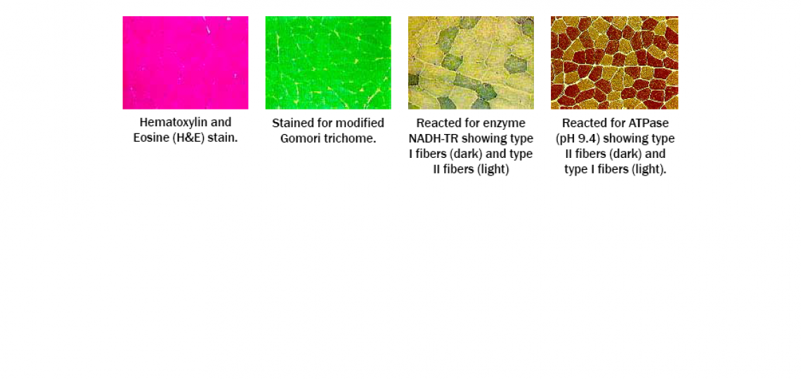
After the muscle specimen is obtained, it is promptly sent to the laboratory. At the laboratory, the specimen is immediately frozen and sectioned. Several routine stains are performed on the muscle specimen. We usually perform H&E, Modified Gomori Trichrome, NADH-TR, and ATPase on the same day of the biopsy so a preliminary report can be given to the treating physician. The preliminary report provides rapid information concerning the presence of inflammation or denervation. Other routine stains performed at our laboratory include succinic dehydrogenase, myophosphorylase, cytochrome C-oxydase, Oil-red-O, periodic acid Schiff (PAS), acid phosphatase, nonspecific esterase, and Congo red. Other histochemical and immunohistochemical reactions will be obtained if necessary. Immunohistochemistry for detection of CD4+, CD8+ cells, macrophages, and other cells are also done in patients with inflammation of muscle or vasculitis. Immunolocalization of dystrophin, complements, and other proteins are also performed when necessary. A final report with accompanying colored photographs representative of the biopsy findings is usually ready about one week after the performance of the muscle biopsy.
Cross-Section of a Normal Muscle


