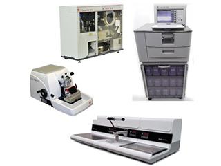
Histology and Pathology Services

- Tissue/cell processing and embedding
- Tissue slide sectioning
- H&E staining
- Special stains including: PAS, ORO, Trichrome, VVG for elastin, Giemsa
- Sectioning of thick tissue curls or core punches for RNA/DNA isolations
Custom Immunohistochemistry (IHC)
- IHC using researcher provided primary antibodies
- IHC using core provided antibodies
- Ki67
- Cleaved PARP-apoptosis (human),
- CD31 (mouse)
- TUNEL assay
- Double antibody IHC
Laser Capture Microdissection
Laser capture microdissection with the Arcturus XT instrument is a way for researchers to enrich a sample for a particular cell type or group of cells. Frozen tissue sections yield better results than FFPE. Contact the lab to see if this technique is appropriate for your project.
Digital Imaging of Stained Slides
Digital scoring of IHC is performed using Perkin Elmer inForm software. inForm can be trained to find virtually any tissue type, structure or tissue subtype. Moreover, inForm can provide area object counts or can be used to automatically assess IHC staining levels on a cell-by-cell and sub-cellular basis, providing extremely accurate and sensitive quantitation.

Tissue Microarray Development
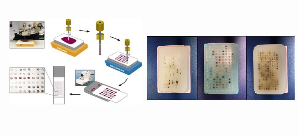
Our core constructs Tissue Microarrays using human or animal model tissues. Creating a TMA from your animal model tissues puts your entire experiment on one slide. All animals, experimental and controls, are stained together under the same conditions. Having a single slide saves money because you section and stain fewer slides, as well as use less antibody and other reagents. Contact the lab to see if TMA construction is appropriate for you animal experiment.
HTAP has built several TMA’s from archival human tissues:
- Prostate arrays
- Multicancer arrays
- Lung cancer array
- Colon cancer array
- Head & Neck Cancer arrays
- Meningioma array
If you don’t see what you need here, contact HTAP to request a TMA build from archived clinical material. HTAP manages an archive that contains formalin fixed paraffin-embedded tissue specimens from a variety of organ/disease types. Approximately 10 to 12 years of clinical, pathological and follow-up data is available for most of these tissue specimens.
Alternatively, TMAs can be built using specimens provided by the investigator and can be designed to address specific questions in the investigator's proposed research. Tissue/cell type and diagnosis for each specimen on TMA is confirmed by a pathologist.
A formal tissue request is required for access to TMA slides.

Human Tissue Acquisition & Distribution
Supported by the Dan L Duncan Comprehensive Cancer Center, human tissue banking takes place routinely at Baylor St. Luke’s Medical Center, the Michael E. DeBakey Veterans Affairs Medical Center and Ben Taub General Hospital. Tissue acquisition also supports clinical trials and research protocols. Specific requests for FFPE, frozen inventory and data management.
Pathology Consultation
One of our experienced pathologists will schedule time to consult with you. They can assist in interpreting your animal model and make recommendations for additional analysis.


