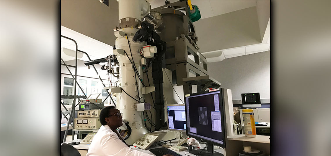
JEOL3200 Electron Microscope
JEM3200FSC (300kV) electron microscope with a field emission gun, an in-column energy filter, a cryo-chuck holding up to three grids, a goniometer tilt range (+/-70° tilts) with dual rotation cartridge, a Gatan K2 Summit direct electron detector, SerialEM for automated single particle and tomography data collection.

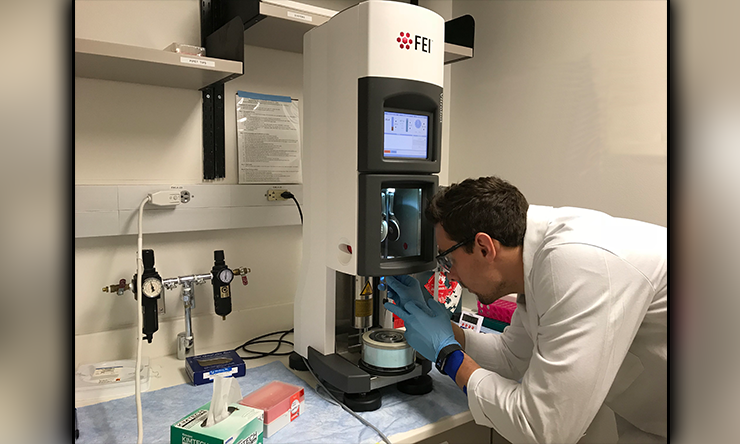
Vitrobot
FEI Vitrobot vitrification robots used for cryo-specimen preparation.

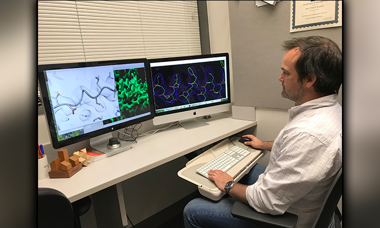
Modeling
Modeling of a large macromolecular assembly at near atomic resolutions from single-particle cryoEM.
About the Core
The CryoEM Core is a state-of-the-art resource for near-atomic resolution 3-D analysis of the structure and dynamics of macromolecules and assemblies, either purified or within cells. This includes the established technique of single particle analysis, whereby images of tens of thousands to millions of isolated macromolecules are reconstructed to produce one or more 3-D structures at resolutions as high as 0.2 nm, as well as in-situ electron cryotomography which permits the 3-D study of cells or regions of cells at resolutions 100x better than optical microscopy. Unlike super-resolution optical microscopy, this is true structural information at 1-5 nm resolution, not just localization.
This later technique is now being extended to extract the structures of macromolecules from within the cellular volume, producing 3-D structure of macromolecules as they are functioning in their native state. For purified complexes, single particle analysis is a direct alternative to X-ray crystallography, and can provide additional information about dynamics and compositional variability, which crystallography cannot access.
Our core is equipped with three high-end electron cryomicroscopes and the associated equipment required to prepare optimal specimens.
We have the equipment and expertise to approach a wide range of structural problems at the 1 nm – 1 micron scale. We can assist with all aspects of CryoEM/ET from optimal specimen preparation through 3-D reconstruction and analysis.
Getting Started
If you plan to make use of the CryoEM Core, we require a brief (no cost) consultation with Dr. Ludtke and/or Dr. Wang for new projects. If you aren't ready for a meeting, but just have questions about the method, feel free to email Dr. Ludtke any time.
To request a new project consultation, register in iLabs, then go to the "Request Services" tab and fill out a "New Project Proposal & Consultation Request" form. This will ask for some details about your specimen, and we will contact you to schedule a meeting. If you are an experienced CryoEM user, and already know precisely what you need, you will still need to fill out the project information form, but we will waive the meeting requirement.
Acknowledging CryoEM Core
For non-cancer-related work:
CryoEM data was collected at the Baylor College of Medicine CryoEM ATC, which includes equipment purchased under support of CPRIT Core Facility Award RP190602.
For cancer-related work:
CryoEM data was collected at the Baylor College of Medicine CryoEM ATC, subsidized by CPRIT Core Facility Award RP190602 which also supported acquisition of CryoEM equipment used in this study.
For any projects making use of the Aquilos (FIB), please add the additional acknowledgement:
FIB milling and CryoEM data collection was performed at the Baylor College of Medicine CryoEM ATC, which includes equipment purchased using CPRIT Core Facility Award RP190602 and additional equipment purchased with a FIB Milling for Cellular CryoET grant (2021FIB-41) from the Arnold and Mabel Beckman Foundation.


