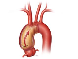
Your aorta is a tube-like structure that resembles a candy cane. The ascending aorta forms the beginning or handle of the cane and originates at the aortic valve. The ascending aorta includes the aortic root and sinuses of Valsalva where the blood supply to your heart, via the coronary arteries, originates. The ascending aorta continues upward to form the aortic arch, where the oxygenated blood supply to your head originates. The aorta continues on down to the descending aorta that further supplies blood to the rest of your body.
What Is an Ascending or Aortic Arch Aneurysm?
An ascending or aortic arch aneurysm refers to ballooning out of the aorta which causes aortic wall weakening. The aortic wall may continue to expand or may remain unchanged, but close surveillance is necessary. As the aortic wall weakens, there is a risk of the wall tearing or dissecting.
What Contributes to the Formation of an Ascending or Aortic Arch Aneurysm?
- Age/degenerative disease of the aortic wall
- Uncontrolled high blood pressure
- Long-term use of tobacco
- Inflammation or swelling of the aorta
- Infection
- History of connective tissue disorders, such as Marfan syndrome, Ehlers-Danlos syndrome, or Loeys-Dietz syndrome
- Trauma
How Can You Tell if You Have an Aneurysm?
Individuals with ascending or aortic arch aneurysms usually do not have symptoms. Ascending and aortic arch aneurysms are commonly found incidentally. It is possible that, as the aneurysm enlarges and compresses surrounding nerves or organs, an individual may experience chest pain, hoarseness or difficulty swallowing.
Patients that experience sudden symptoms such as chest pain, characterized as a tearing sensation, or back pain, nausea, vomiting, a fast heart beat and possibly the feeling of impending doom, may be experiencing a tear or dissection of the aorta. Left untreated this can lead to rupture and is considered an emergent condition that requires immediate intervention.
Diagnostic Evaluation of an Aneurysm or Dissection
Computed tomography (CT) with or without contrast is the most common imaging study used to evaluate your condition. A CT provides valuable information about your aorta, such as the location and size of an aneurysm or dissection. Magnetic resonance imaging (MRI), or magnetic resonance angiography (MRA), is another imaging modality to visualize your aortic aneurysm and vessels. Similar to a CT with contrast, an MRI or MRA provides detailed information about your aorta. Your surgeon will choose the best method for imaging your aorta.
Surgical Repair of Your Aorta
During this operation, the surgeon will make an incision, called a median sternotomy, in your chest and through the breastbone.
In order to perform surgery on your aorta, it is necessary to connect you to a heart-lung bypass machine. This machine takes over the work of the heart circulating oxygenated blood throughout the body. During your surgery, the diseased portion of your aorta will be replaced with a Dacron® graft. If you have an arch aneurysm, it is possible that you may also require bypass grafts to replace vessels that supply blood to your head.
While repairing your aorta, your surgeon will inspect your aortic valve to determine whether it can be spared or may need replacement, which is done at the same time as your aneurysm surgery. Please, see the separate insert about valve replacement for more information.
Recovery
It will take you approximately two to three months to fully recover from undergoing ascending aortic aneurysm/dissection or arch surgery. You should plan to be away from work, getting your full strength back, for six to eight weeks. Your surgeon will advise you on post-operative restrictions and when it’s safe to drive again. Participating in a cardiac rehabilitation program will be beneficial in your recovery. Make sure to ask about this before you are discharged from the hospital and make arrangements close to your home in advance.


 Credit
